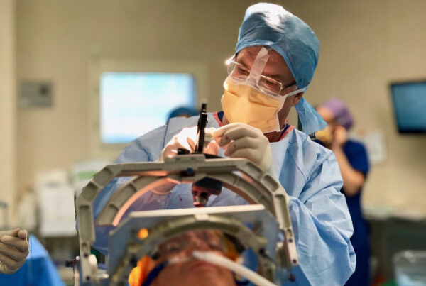
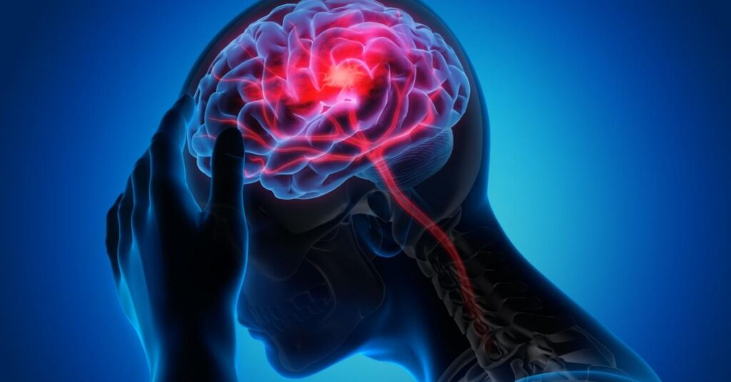
In neurosurgery, brain tumor removal is one of the most complex and precise procedures, given the sensitivity of brain tissue and its connection to vital functions such as movement, speech, and memory.
Brain tumor removal is a surgical procedure aimed at removing the tumor to the maximum extent possible while preserving the brain’s functional capabilities.
With medical advancements, new techniques have emerged that balance safety and complete tumor resection.
With tremendous progress in surgical techniques, these operations have become safer and more effective, giving patients better chances of recovery and a satisfactory quality of life.
What are Brain Tumors?
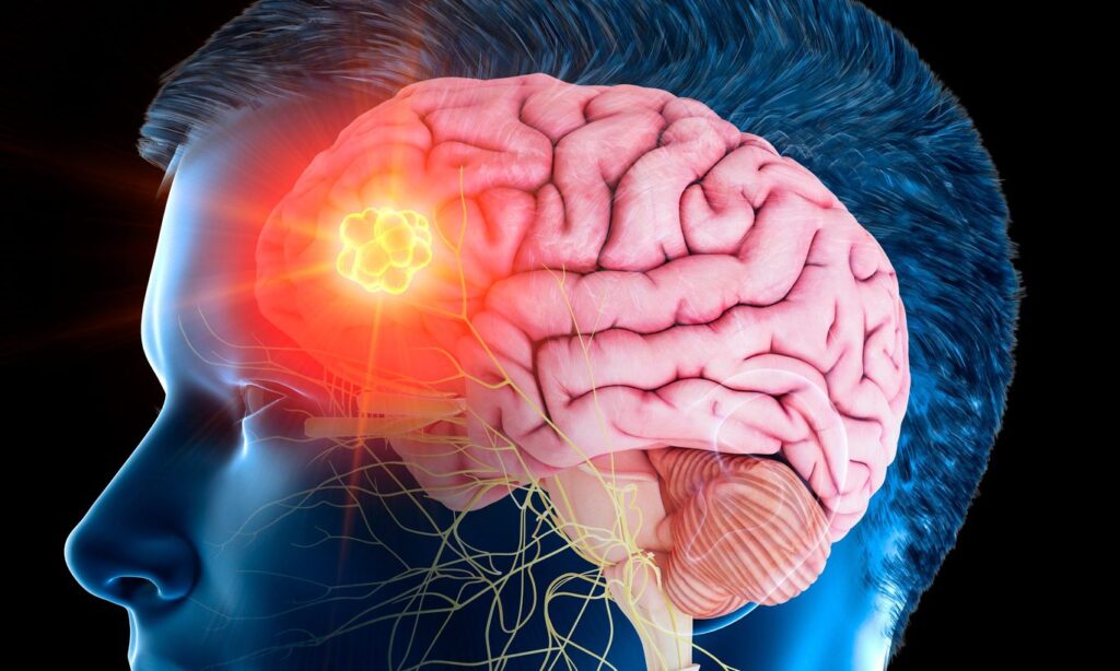
Brain tumors are abnormal growths of cells inside the brain or its surrounding membranes.
Common symptoms include chronic headaches, seizures, vision disturbances, and impaired motor or cognitive functions. They are divided into two main types:
- Benign Tumors: Grow slowly and do not spread, but they can cause complications due to pressure on brain tissue.
- Malignant Tumors: Grow rapidly and may spread to other areas, such as Glioblastoma.
Modern Brain Tumor Removal Procedures
Brain tumor removal surgeries no longer carry the significant risks associated with them in the past.
Thanks to modern techniques and specialized medical teams, such as those at Liva Hospital, these operations have become a real gateway to hope for patients.
Early diagnosis and choosing the appropriate medical center are the most influential factors in treatment success.
Initially, brain surgeons collaborate with multidisciplinary medical teams (oncology, radiology, neurophysiology, etc.) for precise planning.
Pre-surgical neuro-mapping techniques such as functional MRI (fMRI) and neuroendoscopy are used to identify and avoid sensitive areas during resection.
Advanced Guidance Techniques: Fluorescein and Spectroscopy
Fluorescence-guided surgery (5-ALA) gives a special luminescence to tumor tissues under the microscope, helping the surgeon remove more than 65% of the tumor and enhancing progression-free survival for up to six months.
Raman spectroscopy also helps distinguish tumor tissue from healthy cells during the operation instantly with high accuracy.
Keyhole Surgery and Endoscopic Surgery
Microsurgical or endoscopic surgery through small incisions (Keyhole/endoscopic) reduces the size of wounds and speeds up recovery.
One of the most prominent examples is the removal of pituitary tumors through the nose (endoscopic endonasal), where the complete resection rate has reached about 90% even for large tumors.
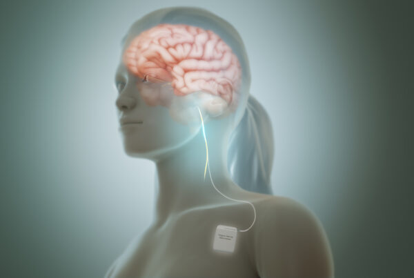
Awake Craniotomy
In this surgery, the patient remains conscious while the tumor is removed in sensitive areas such as the language or motor center.
This method helps in immediate neuro-mapping to avoid damage to vital functions, with better recovery outcomes and higher total resection rates.
Deep Tools: Tubular Retractors
Techniques like BrainPath provide a narrow path to access deep-seated tumors with better pressure distribution on tissues, reducing damage.
Research has indicated that the complete resection rate reaches about 80%, with higher safety compared to traditional surgery.
Machine Learning and Spectral Analytics
Deep learning techniques are used to improve the quality of intraoperative images, such as restoring iMRI images and highlighting suspicious margins.
Preliminary studies achieved a classification accuracy of up to 96% in cellular differentiation of tumor tissues.
Conclusion
In short, brain tumor removal has seen amazing developments in recent years.
From precise planning to smart tools and complementary interventions, the ultimate goal is to achieve maximum resection with patient safety.
Techniques such as iMRI, neuronavigation, fluorescence-guided surgery, endoscopic surgery, and awake craniotomy represent a promising future for more successful and safer treatment.
The future of operations, thanks to artificial intelligence techniques, is bright towards unprecedented accuracy.
Liva Hospital is one of the advanced centers that rely on modern techniques mentioned such as iMRI, neuronavigation, endoscopic surgery, and awake craniotomy.
A multidisciplinary team works there according to international medical protocols to achieve the highest levels of safety and reduce complication rates.
Frequently Asked Questions (FAQ)
What is the difference between iMRI and traditional surgery?
iMRI allows for live imaging during the operation to identify residual tumor, increasing the likelihood of complete removal and reducing the need for re-intervention.
Is awake craniotomy painful?
Local anesthesia and psychological preparation are used, so the patient does not feel severe pain; they are only asked to cooperate during neuro-mapping.
What is the 5-ALA technique and how safe is it?
It is a substance used before surgery that accumulates in tumor cells and glows under the microscope. It is relatively safe with minor side effects such as skin sensitivity to sunlight.
Can machine learning be used in all hospitals?
Currently, no. This technology is still under development and requires special equipment and additional research, but it is promising and may become a standard in the future.
Is Liva Hospital a reliable option for brain tumor treatment?
Yes, Liva Hospital adopts modern techniques and includes multidisciplinary teams, which enhances the quality of care and patient trust.
Is recovery possible after complete resection?
The results depend on the type of tumor, its location, and the extent of its removal, but modern techniques clearly contribute to improving recovery rates and the speed of neurological recovery.